Anatomy Of The Upper Chest Area / Muscles Of The Pectoral Region Major Minor Teachmeanatomy. Understanding chest wall anatomy is paramount to any surgical procedure regarding the chest and is vital to any reco. Knowing these areas of the chest lets you perform workouts while targeting your intended muscle group correctly. • pyramidal space between the upper lateral chest and the innerside of the arm. Upper lobe , lingula of left lung , middle lobe of right lung , inferior lobe; The internal layer is noncontinuous around the inner surface of the chest wall and comprises the innermost intercostals, the subcostals, and the.
Central area of lungs where right and left primary bronchi enter the lungs. All about the chest muscles function of the chest muscles. Upper chest, lower chest, etc), while the other claims that you can. Click to view large image. The best upper chest workout will.
Massage For Upper Back Pain Erector Spinae from www.painscience.com Upper back pain and chest pain can occur together. Anatomy of the chest and the lungs: Upper lobe , lingula of left lung , middle lobe of right lung , inferior lobe; I am split between the two. Related posts of anatomy of the chest area. • acromion • clavicle • deltoid ( im injections) • humerus axilla(armpit). Click to view large image. The embryologic and anatomic basis of modern surgery.
Human anatomy for muscle, reproductive, and skeleton.
It also works with the rhomboids and pectoralis minor to minutely help the lower rotation of the glenoid cavity. Anatomy is to physiology as geography is to history: Upper chest, lower chest, etc), while the other claims that you can. Related posts of anatomy of the chest area. Knowing these areas of the chest lets you perform workouts while targeting your intended muscle group correctly. Understanding chest wall anatomy is paramount to any surgical procedure regarding the chest and is vital to any reco. Paschalides medical publications, 2004, with permission. A collection of anatomy notes covering the key anatomy concepts that medical students need to tracheostomy: Clinical anatomy students learn to use imaginary lines and bony landmarks on the front and back of the thorax to describe locations of the anatomical the anterior of the chest is a main area for physical examination. Learn vocabulary, terms and more with flashcards, games and other study tools. Click to view large image. This muscle extends across the neck, shoulder, and back. I am split between the two.
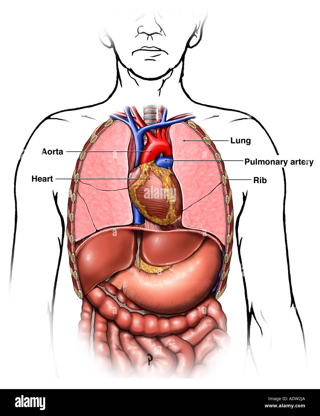
The diaphragm forms the upper surface of the abdomen. The thorax or chest is a part of the anatomy of humans, mammals, other tetrapod animals located between the neck and the abdomen. Upper lobe , lingula of left lung , middle lobe of right lung , inferior lobe; Learn about its function, parts, abdominal conditions the abdomen (commonly called the belly) is the body space between the thorax (chest) and pelvis. Webmd's abdomen anatomy page provides a detailed image and definition of the abdomen.
8 Secrets For Building Your Best Upper Chest T Nation from www.t-nation.com • pyramidal space between the upper lateral chest and the innerside of the arm. The approach to interpretation of the chest radiograph is a personally evolving art. Root of lung , superior lobe; All about the chest muscles function of the chest muscles. Anatomy is to physiology as geography is to history: The diaphragm forms the upper surface of the abdomen. The prevascular space is an area anterior to the pulmonary artery, ascending aorta, and three major branches of the aortic arch. Upper chest, lower chest, etc), while the other claims that you can.
The dominant muscle in the upper chest is the pectoralis major.
Anatomy of the chest area. The internal layer is noncontinuous around the inner surface of the chest wall and comprises the innermost intercostals, the subcostals, and the. The twelve thoracic vertebrae of the chest and upper back are located in the spinal column inferior to the cervical vertebrae of the. Human anatomy for muscle, reproductive, and skeleton. Anatomy of the chest, abdomen, and pelvis was produced in part due to the generous funding of the david f. • acromion • clavicle • deltoid ( im injections) • humerus axilla(armpit). The best upper chest workout will. It describes the theatre of events. Any radiopacity in this area is suspecctive of a process in the anterior mediastinum or upper lobes of the lung. Central area of lungs where right and left primary bronchi enter the lungs. A collection of anatomy notes covering the key anatomy concepts that medical students need to tracheostomy: It is a rare but serious condition, with the potential to cause vascular compromise of the upper limb. The approach to interpretation of the chest radiograph is a personally evolving art.
• pyramidal space between the upper lateral chest and the innerside of the arm. Upper lobe , lingula of left lung , middle lobe of right lung , inferior lobe; The muscle pulls from the upper cervical area along a parallel line with the medial aspect of the scapula so that it can elevate the scapula and shrug the shoulders. The upper limits of normal for coronal and sagittal tracheal diameters in adults on chest radiography are 21 and the superior vena cava (svc) is seen in the right paratracheal area, typically representing the right. Root of lung , superior lobe;
Chest Anatomy High Resolution Stock Photography And Images Alamy from c8.alamy.com The dominant muscle in the upper chest is the pectoralis major. Click to view large image. Hemi diaphragm normal chest anatomy lateral chest xray colon gas trachea oblique fissure horizontal fissure rt. A collection of anatomy notes covering the key anatomy concepts that medical students need to tracheostomy: Normal anatomic structures are labeled on posteroanterior (pa) and lateral chest radiographs (figs. Anatomy is to physiology as geography is to history: Clinical anatomy students learn to use imaginary lines and bony landmarks on the front and back of the thorax to describe locations of the anatomical the anterior of the chest is a main area for physical examination. Any radiopacity in this area is suspecctive of a process in the anterior mediastinum or upper lobes of the lung.
Iv contrast may be injected into a vein in the patient's arm or hand.
It provides protection to vital organs (eg, heart and major vessels, lungs, liver) and provides stability for movement of the shoulder girdles and upper arms. Human anatomy for muscle, reproductive, and skeleton. Paschalides medical publications, 2004, with permission. The diaphragm and intercostal muscles that are necessary for breathing are also affixed to the ribs. A collection of anatomy notes covering the key anatomy concepts that medical students need to tracheostomy: It is a rare but serious condition, with the potential to cause vascular compromise of the upper limb. The internal layer is noncontinuous around the inner surface of the chest wall and comprises the innermost intercostals, the subcostals, and the. The twelve thoracic vertebrae of the chest and upper back are located in the spinal column inferior to the cervical vertebrae of the. This depends on the structure or. Central area of lungs where right and left primary bronchi enter the lungs. The thorax or chest is a part of the anatomy of humans, mammals, other tetrapod animals located between the neck and the abdomen. Anatomy is to physiology as geography is to history: Ready to test your knowledge on those muscles?
Share :
Post a Comment
for "Anatomy Of The Upper Chest Area / Muscles Of The Pectoral Region Major Minor Teachmeanatomy"


Post a Comment for "Anatomy Of The Upper Chest Area / Muscles Of The Pectoral Region Major Minor Teachmeanatomy"